Dindigul Hospital
For Emergency: Madurai: 0452 2501033, Dindigul: 0451 2444444
Interventional Radiology

Interventional Radiology

The Department of Interventional Radiology at Vadamalayan Hospitals is dedicated to the development and application of imaging to guide minimally invasive techniques utilized in the diagnosis and treatment of a wide variety of conditions. A medical sub-speciality, Interventional Radiology utilizes minimally-invasive image-guided technique to diagnose and treat nearly every organ system. The objective of Interventional radiology is to minimize risk to the patient and improve health outcomes.

Procedure

The Interventional Radiology procedure offers the most in-depth knowledge of the minimally invasive treatments available using needles, wires and catheters to deliver treatments through a pinhole with minimum morbidity and less recovery time compared to conventional surgical procedures. The Interventional radiologists use X-Ray, CT and other imaging to advance instruments in the body, such as in an artery to treat the disease at the source. Many conditions that once required surgery can be treated less invasively by Interventional radiologists. These treatments offer less risk, less pain and less recovery time compared to open surgery.

Interventional Radiologists are image-guided therapy clinicians who are specially trained in image interpretation and minimally invasive treatments of a wide variety of conditions across multiple specialities. Interventional Radiologists can deliver treatments for various-cancers, fibroids, back and joint pain, varicose veins, arterial diseases, kidney and bile duct disease to name a few.

Interventional Techniques

With the enhancement of technology, the interventional radiology procedures are a boon. Various kinds of engineering are used in the medical field of interventional radiology for imaging the internal parts of a human body. Some of the techniques are mentioned below.

  • Embolisation
  • Chemoembolization
  • Ablation
  • Balloons and stents
  • Needle biopsy and drainage
  • Other techniques
Embolisation

A Catheter-based technique using coils, particles or glue to block tumour vessels or acute bleeding.
When a tumour grows in our body, it requires a constant flow of blood. The blood keeps the tumour alive. In case the blood supply stops, then the tumour does not get any nourishment and slowly dies.

Embolization is a unique technique that allows interventional radiology specialists to nip the infection in the bud. The radiologist tries to cut the source of blood carefully so that the tumour is unable to grow any further. In the process of Embolisation, there is a minimum amount of interference to the other organs or blood vessels.

The process of embolization requires a massive amount of liquid along with minute particles flushed into the body. This way, the blood flow in a particular area gets blocked due to the minute particles gradually stopping the source of nourishment for the tumour. The process of embolization is beneficial for patients suffering from a tumour in their lungs, liver, kidneys, or bones.

There are different procedures in which embolization can be done in interventional radiology. Before starting the process, the interventional radiologist gives local anaesthesia to the patient. As the procedure may take more than an hour to complete, the application of anaesthesia ensures that the patient doesn’t make any kind of movement and the accurate diagnosis of the tumour can be obtained.

Moving ahead, the radiologist punctures the thighs of the patient with a minute needle to advance catheters and guidewires, a threadlike cluster of plastic tubes, in the arteries of the patient. After this, a contrast medium dye will be injected into the catheter to get a clear view of the imaging.

Once a clear view is obtained, the interventional radiologist will inject embolic particles carefully by using a microcatheter. The particles injected are usually 0.5 mm in size and contain a considerable dose of chemotherapy or yttrium radiation to cut off the flow of the blood to the tumour.

There are particular side effects noted among the patients who underwent this process, such as nausea or pain. However, consuming a medically approved painkiller shall provide certain relief.

Chemoembolization

Same as above but using local targeted high dose chemotherapy to treat tumours.
Chemoembolization is another reformed process of embolization. In the process of chemoembolization, the interventional radiologist will place a minute-catheter in the blood vessel and inject it into the arteries. These arteries supply blood to the liver.

The process of chemoembolization is similar to that of a cardiac angiogram. The drugs used for chemotherapy are injected into the catheter along with blood vessels, which shall restrict the area of a tumour getting spread. The entire process involves the functioning of a highly effective anti-tumoral drug in the infected regions fighting the tumour from within. The double attack on the tumour shall stop the spread of it.

Various kinds of tumour-like hepatoma or primary liver cancer metastasise and spread to the liver from colon cancer, carcinoid, ocular melanoma, sarcomas or any other kind of tumour in another part of the body.

Ablation

Needle or catheter-based technique using thermal energy forms like radiofrequency, microwave or cryo technique to treat tumours or varicose veins.
Ablation is also popularly known as catheter ablation. In the process of ablation, either heat or extreme cold is applied to the infected area. The heat radiation is known as radiofrequency ablation, and the freezing radiation is called cryoablation.

The treatment has dire effects on the body, like abnormal braking circuits in the heart. It sometimes also destroys areas near the heart muscles triggering symptoms of arrhythmias.

The process of ablation is similar to that of the interventional radiology procedures of electrophysiology (EP) study, which is done to detect the abnormal heart rhythm. The ablation process is beneficial for patients who have been detected with the abnormalities early. The entire process requires 8-10 weeks.

Balloons and stents

Techniques to open up a blocked tube like an artery, vein, bile duct, ureter, colon or oesophagus.
Another unique interventional radiology procedure is the Balloon Angioplasty and Stents, where a long catheter with balloon ends are used to remove the plaque from the affected area. This procedure is performed in the cardiac catheterization laboratory, also known as Cath lab. The balloons and stents are the best tools that can be used in the treatment of coronary artery disease or CAD. Usually, CAD cannot be managed by medicines. Therefore, balloons and stents technique works best for CAD.

The process involves a twist in the old fashion angioplasty. A long tube or catheter is designed with balloon-shaped tips. The balloons are inflated at the blockage site in the artery. This way, the plaque gets flattened, or even compressed, against the walls of the artery. This kind of angioplasty is also known as percutaneous transluminal coronary angioplasty or PTCA.

The balloon angioplasty is quite beneficial as it can be used to open the vessels, which has become narrow or has shrunk down in many parts of the human body. The stent is a minute mesh-like device used in modern interventional radiology. It is made up of substances with metallic nature. The stent acts as a support or scaffold in the interventional radiology procedures.

Usually, the stent is placed inside the coronary artery, which aids in keeping the blood vessel open. In this way, the stents help in improving the blood flow to the muscles of the heart, which eventually reduces the pain of angina.

The stents are usually built with balloon-like fixtures at the end of the tube or catheter. These kinds of angioplasty are found in more than 80 per cent of patients who get balloon angioplasty. Doctors use stents like this for body parts other than the heart as well. Stents can also be used in the medical procedure involving carotid arteries in the neck or the arteries in the aorta along with the peripheral arteries in the legs.

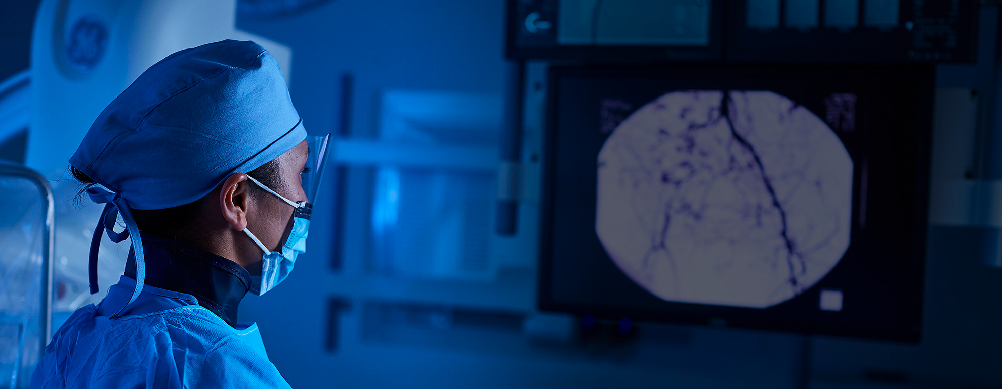
Doctors usually suggest their patients to avoid drinking or eating after midnight prior to the day these tests are conducted. In case a patient has diabetes, then it is essential to consult the doctor before undergoing the procedure; otherwise, it might affect the blood pressure of the patient. During the procedure, the doctor attaches a catheter that goes into the artery inside the heart. A video monitor is used to see the inside of the body. Once the doctor has detected the blocked area, a dye is injected into it. This helps the doctor to get a snapshot of the coronary arteries. This method is also known as a coronary angiogram.

Once the blocked area is detected, the doctor ties a thread, also known as guidewire around the artery. After that, the balloon tip is inflated in that area, which subsequently presses against the plaque, causing compression against the artery walls. The inflation and deflation of the balloon are done several times to compress the affected area. The stent also contains medicine, which avoids further spreading of the plaque.

Needle biopsy and drainage

Core biopsy for pathological sampling or for draining thoraco-abdominal collections and abscesses.
Needle biopsy is considered to be one of the most effective ways to collect samples of the cell or tissue from the body for laboratory testing. Usually, a common needle biopsy comes under two ways:

fine-needle aspiration
core needle biopsy
Needle biopsy is conducted to extract all the tissue and fluid samples from different parts of the body that includes the muscles and all other organs. This type of dialysis is conducted to treat diseases or disorders. Needle biopsy enables the chances of getting an accurate result.

In case a sudden lump or mass is found on the body, and the doctor is unable to detect the source or the reason for the lump, then the doctor suggests to get a needle biopsy done. A needle biopsy may also be suggested by the doctor if an infection or inflammation of any kind is detected at a particular part of the body.

Patients should consult the doctors in case there is any liquid oozing from the place biopsy is done.

Other techniques

Clot retrieval or lysis, caval filters, aneurysm stents, sclerotherapy etc.
The lysis test is conducted to see how RBC (or red blood cells) absorb complement components with the help of serum at a low ionic rate.

The caval filter technique is another unusual interventional radiology procedure in which a filter is attached in a large vein situated at the abdomen that returns blood from the lower body to the heart.

Most of the time, these blood clots can be noticed in the veins and pelvis, that randomly reaches to the lungs, causing a blockage or pulmonary embolism. Caval filters are known to reduce the vulnerabilities of pulmonary embolism. This helps in trapping the clots and protecting it from entering in the heart or lungs. These are some of the practical techniques that form a part of interventional radiology procedures.

Advantages
  • Day case or short admission procedures
  • Less / no pain
  • More cosmetic – pinhole/spot dressing surgery
  • Less morbidity/complications
  • More rapid return to work
  • Does not close the door for conventional surgeries
  • Can be combined with other treatments like chemotherapy
  • Conditions that can be treated include
    Most uterine fibroids
  • Rest pain and claudication
  • Ischaemic diabetic foot
    Varicose veins
  • Male varicocele and pelvic congestion syndrome
  • Benign prostatic hyperplasia
  • Recurrent haemoptysis, GI bleeds
  • Back pain
  • Curative treatment for small kidney, lung or liver tumours
  • Local control for larger tumours
  • Venous access and ports
    Renal dialysis access
    Bile duct and ureteric blocks
    Complications of portal hypertension

Our Doctors (Madurai)

  • Dr.M.Narasimman M.D AIMS (FIVR).,

    Interventional Radiologist

    Madurai / Dindigul